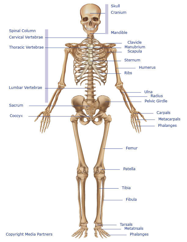
Pic Structure Of Human With All Muscles And Bones Name : Human Skeletal System : Of all 24 ribs, the first seven pairs are often labeled as 'true.'.
byAdmin-
0
Pic Structure Of Human With All Muscles And Bones Name : Human Skeletal System : Of all 24 ribs, the first seven pairs are often labeled as 'true.'.. Bone anatomy arm 12 photos of the bone anatomy arm arm bone anatomy quiz, bone anatomy arm, bone. Tendons connect the knee bones to the leg muscles that move the knee joint. The skull from a 3⁄4 frontal view frontal b. The bony bumps (or protrusions) seen and felt on the ankle have their own names: The biceps is the chief flexors of the forearm.
Neck anatomy pictures bones, muscles, nerves. The purpose of the spine is to support the body so that we can stand upright. This is a table of skeletal muscles of the human anatomy. Tendons are tough bands of dense regular connective tissue whose strong collagen fibers firmly attach muscles to bones. Acetabular shell which will be inserted into pelvis.
Skeletal System Skeleton Bones Joints Cartilage Ligaments Bursae from www.healthpages.org The biceps is the chief flexors of the forearm. Just need a glimpse, leave your valuable advice let us know , and subscribe us! Related posts of human bones and body images with name diagram labeling the structure of a bone. An illustrated anatomy head and neck illustration 1 parietal b. Order personalized gifts to keep your loved ones close forever. It consists of calcified material, which, coincidentally, is also known by the same name 'bone.' from a scientific view, a bone is a substance that forms the skeleton of the body chiefly made of calcium phosphate and calcium carbonate, and serves as a storage area. We are pleased to provide you with the picture named labelled diagram of the muscles in the human body.we hope this picture labelled diagram of the muscles in the human body can help you study and research. This framework consists of many individual bones and cartilages.there also are bands of fibrous connective tissue—the ligaments and the tendons—in intimate relationship with the parts of the skeleton.
Molly smith dipcnm, mbant • reviewer:
Diagram labeling the structure of a bone 12 photos of the diagram labeling the structure of a bone , bone Muscles of the back anatomy muscles of the back anatomy isolated on white background. Just need a glimpse, leave your valuable advice let us know , and subscribe us! The tarsal bones are found near the. Secondarily, it protects the spinal cord (which is the extension of the brain) and all of the nerves that branch from the spinal cord. Tendons connect the knee bones to the leg muscles that move the knee joint. Human skeleton, the internal skeleton that serves as a framework for the body. The triceps is an extensor muscle of the elbow joint. It is composed of 300 bones at birth, but later decreases to 80 bones in the axial skeleton and 126 bones in the appendicular skeleton. The human rib cage is made up of 12 paired rib bones; It allows the upper limb to have a wide array of movements. See human body anatomy stock video clips. Bone anatomy arm 12 photos of the bone anatomy arm arm bone anatomy quiz, bone anatomy arm, bone.
Customize a beautiful piece of jewelry with your favorite stones or an engraving. Bone anatomy arm 12 photos of the bone anatomy arm arm bone anatomy quiz, bone anatomy arm, bone. Browse 1,010 hip bone stock photos and images available, or search for hip bone icon or hip bone 3d to find more great stock photos and pictures. This article is concerned primarily with the gross structure and the function of the skeleton of the normal. One row connects with the ends of the bones in the forearm—the radius and ulna.
The Human Organ Systems Human Anatomy And Physiology Lab Bsb 141 from s3-us-west-2.amazonaws.com September 23, 2019 may 6, 2015 by dr. It is composed of 300 bones at birth, but later decreases to 80 bones in the axial skeleton and 126 bones in the appendicular skeleton. Our latest youtube film is ready to run. Customize a beautiful piece of jewelry with your favorite stones or an engraving. Dimitrios mytilinaios md, phd last reviewed: Neck anatomy pictures bones, muscles, nerves. The shoulder joint is one of the most movable joints in the human body. It is comprised of many bones, formed by intramembranous ossification, which are joined together by sutures (fibrous joints).
Secondarily, it protects the spinal cord (which is the extension of the brain) and all of the nerves that branch from the spinal cord.
Take the following quiz t. Ligaments join the knee bones and provide stability to the knee: Each are symmetrically paired on a right and left side. The purpose of the spine is to support the body so that we can stand upright. Gross anatomy of a skeletal muscle. Secondarily, it protects the spinal cord (which is the extension of the brain) and all of the nerves that branch from the spinal cord. Anatomy of the hand and wrist: The wrist links the hand to the forearm. May 31, 2021 reading time: These bones provide structure and protection and facilitate motion. , the shell has a porous surface which allows it to integrate with surrounding. Skeletal muscle derives its name from the fact that these muscles always connect to the skeleton in at least one place. 11 f bones and muscles:
Take the following quiz t. The carpal bones are arranged in 2 interrelated rows. Human arms anatomy diagram, showing bones and muscles while flex. Anatomy of the hand and wrist: An illustrated anatomy head and neck illustration 1 parietal b.
1 from The human rib cage is made up of 12 paired rib bones; Cartilage is more flexible than bone but stiffer than muscle. Muscle anatomy reference charts author: Related posts of human bones and body images with name diagram labeling the structure of a bone. Almost every muscle constitutes one part of a pair of identical bilateral muscles, found on both sides, resulting in approximately 320 pairs of muscles, as presented in this article. The triceps is an extensor muscle of the elbow joint. Skull system spinal column icon spine osteoporosis lunbar spine discs spine vertebrae medical skeleton named anatomy of spine spine disc thoracic anatomy diseases of the spine. May 31, 2021 reading time:
See human body anatomy stock video clips.
Ligaments join the knee bones and provide stability to the knee: Human skeleton, the internal skeleton that serves as a framework for the body. The triceps is an extensor muscle of the elbow joint. The bones of the skeletal system act as attachment points for the skeletal muscles of the body. This article is concerned primarily with the gross structure and the function of the skeleton of the normal. Secondarily, it protects the spinal cord (which is the extension of the brain) and all of the nerves that branch from the spinal cord. This framework consists of many individual bones and cartilages.there also are bands of fibrous connective tissue—the ligaments and the tendons—in intimate relationship with the parts of the skeleton. Almost every muscle constitutes one part of a pair of identical bilateral muscles, found on both sides, resulting in approximately 320 pairs of muscles, as presented in this article. See human body anatomy stock video clips. Human arms anatomy diagram, showing bones and muscles while flex human arms anatomy diagram, showing bones and muscles while flexing. The bones of the appendicular skeleton provide support and flexibility at the joints and anchor the muscles that move the limbs. There are around 650 skeletal muscles within the typical human body. Muscle anatomy reference charts author: